
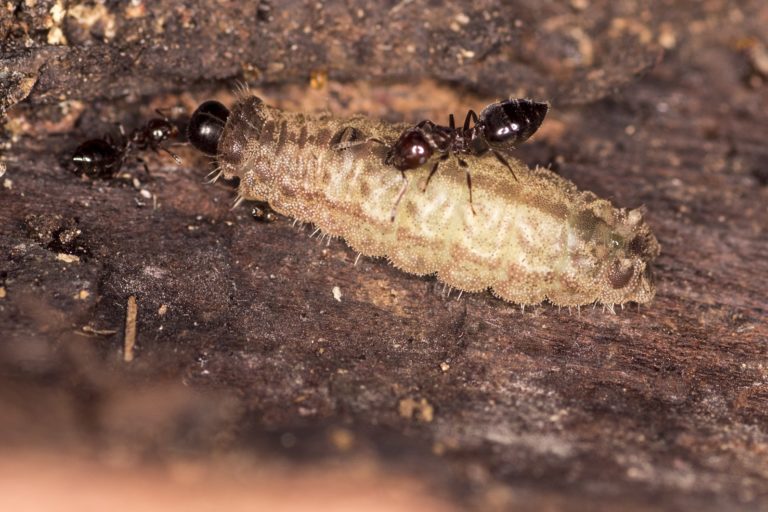
- Butterflies belonging to the family Lycaenidae are well-known for their ant-associations. This myrmecophily, literally “ant love”, is so evolved that the larvae of many species have developed specific body parts to enable this. Ant presence, in return, provides caterpillars protection from predators.
- Using micro-CT scans, scientists unravelled the internal structure of some of these specialised organs in caterpillars of the lilac silverline, a lycaenid endemic to India. They detailed the morphology of dew patches and nectar glands that produce sugary solutions to attract ants, as well as tactile glands that produce ant-attracting chemicals.
- They also found that the caterpillars sport unusually thick skin, including thick plates at their heads to protect them from ants. Such detailed structural data are often the first step to better understanding butterfly-ant associations, and the evolutionary arms race between these insects.
Some butterflies go to any lengths to make ant-friends. Their caterpillars have evolved button-like spots on their backs to spout sugary rewards to attract ants; a special gland in their abdomen concocts nectar to bait ants, and tufted tubular organs help sense ant-chemicals or pheromones. Now, scientists have unravelled the precise internal anatomy of some of these less-known organs, bringing science one step closer to decoding the complex, inter-taxa associations between butterflies and ants.
There’s a word for the ant-friendships that some butterflies display: myrmecophily (literally, ‘ant love’). Most lycaenids, or butterflies belonging to the family Lycaenidae (almost 6,000 species worldwide), are myrmecophiles. And they show their love in myriad ways. Caterpillars of most species have small pores on their skin – called pore cupola – that secrete substances to pacify ants that may otherwise attack them. Others have dew patches, small button-like spots on their backs that ooze a thick sugary fluid to attract ants. Tactile or tentacle organs (that resemble sea anemones, except that their sensitive tentacle-like tufts are sparse) that pop out of their bodies secrete chemicals that also attract ants, and even alert them if the caterpillars are alarmed. Some species have exceptionally thick skin to tolerate ant aggression. Many also have a nectar gland (called the Newcomer’s gland), that provides sugar-rich nectar to ants through an opening on the skin. These features occur in various permutations and combinations: while some lycaenids sport only pore cupola, others display the entire suite of specialised organs.
These are costly features to invest in, but the trouble’s worth it: ant presence provides protection to caterpillars from predators. Such fascinating butterfly-ant associations range from mutualism (where both associates benefit from the interactions) to parasitism (where one benefits at the expense of the other), and from facultative (where ants tend to lycaenid larvae only occasionally and the caterpillars do not depend on them for survival) to obligate (where the larvae are entirely dependent on ants; such as some caterpillars that feed exclusively on food regurgitated to them by ants).
But while we do know about these associations, and that specialised organs facilitate them, an in-depth understanding of these organs is limited. What do their internal structures look like, and how do they function? The lilac silverline (Apharitis lilacinus) – a lycaenid endemic to India that was rediscovered in 2013 in Bengaluru after more than a century – should be an ideal candidate to study and find out, thought Dipendra Nath Basu and Krushnamegh Kunte, scientists at the National Centre for Biological Sciences (NCBS) in Bengaluru.
That’s because recent observations reveal that its caterpillars share an obligate relationship with a cocktail ant, Crematogaster hodgsoni. Female lilac silverlines deposit eggs near the nests of these ants. Ants then attend the butterfly babies at all times: from the second caterpillars emerge, till the moment the pupae take wing as adults. So, armed with special permission from the Karnataka Forest Department (lilac silverlines are protected under Schedule II of the Wildlife Protection Act, 1972), Basu and Kunte collected two caterpillars from the outskirts of Bengaluru, where the only known population of lilac silverlines survive.
Imaging a one-centimetre caterpillar
To study the caterpillars’ specialised body parts, the team used one of the latest methods available: microtomography, or micro-CT. This imaging technique works much like a human CAT scan: it uses tissue-penetrating x-rays to obtain cross-sectional, detailed images. Following several steps to preserve their caterpillars including soaking them in ethanol, the team mounted their specimens on the microtomographer at the NCBS Electron Microscopy Facility. The specialised organs then swung into full view.
A pair of dew patches, like little spherical pots, were located on the caterpillar’s back. In caterpillar morphology, this translates to the top, and front portion, of the larva’s abdomen, as seen in the following figure.

A cross-sectional view of this gland appears almost like that of a volcano: a shallow crater-like depression at the top accommodates sugary liquids temporarily. These liquids are secreted by a string of apocrine and eccrine glands (the tiny red-coloured lobules in the figure above; in humans, these function as sweat glands on the skin) attached to the base of the depression, said Basu, who collected and analysed the micro-CT data. The dew patch opens and closes in a “lasso bag” or drawstring motion, as the caterpillar’s dorsoventral retractor muscles constrict and relax, write the scientists in their study published in Scientific Reports. Once closed, special elongated scales protect the dew patches.
The nectar gland, on the other hand, is located at the other end of the caterpillar’s stomach (closer to its posterior). The nectar travels via a duct and opens onto the skin through a small cleft as seen in the figure below.

When ants palpate the area, the caterpillar’s accessory longitudinal muscles should tighten and possibly push the nectar out; several muscles including the sphincter control this process, suggest the scientists. Ants don’t always get to the nectar: caterpillars can, in fact, suck their nectar back in (considering the high energy requirements of making the nectar), and the last abdominal ganglion (structures containing nerve and tissue cells) probably control this balance, they add.
Finally, the pair of tactile organs – much like a sparsely-tentacled anemone – are located in the posterior segment of the caterpillar and pop out of its body when ants palpate the area. Ventral muscles and haemolymph (the blood equivalent in insects) pressure could be controlling its movement.
Additionally, the team found that lilac silverline caterpillars have very thick and convoluted skin, as well as thick headplates, to protect them from ant aggression. Also, small foreguts. That’s unusual because most caterpillars need wide foreguts to digest the foliage they eat, said Basu. This suggests that trophallaxis – the phenomenon where larvae depend on food regurgitated by ants – could be at work. Detailed studies would be needed to explore this, as well as the other qualitative, quantitative and comparative aspects (such as the lack of ant-associated features in the pupa, which could suggest that pupae maintain their ant associations purely via chemical signals) that the study opens up, he added.

Why are such studies important?
These mechanisms detailed in the study indicate how caterpillars control how much they reward the ants and when, said Kunte, associate professor at NCBS.
“The fine control over the rewards potentially helps them minimise their investments in keeping the ants engaged, at the same time reaping as many benefits from this association as possible,” he wrote in an email to Mongabay-India.
This understanding of the morphology and behaviour of lycaenids can help us understand the interactions and needs an organism has for survival, commented Steen Dupont, a scientist at London’s Natural History Museum who did not participate in the study. Dupont’s study of moth butterfly caterpillars in 2016 showed that fine microscopic structures on their outer skins provide a tough but flexible protection from weaver ants.
“In the lilac silverline the foregut is reduced and we understand that food is primarily through interaction with ants. In this scenario the morphology of the caterpillar tells us that if preservation of the butterfly is the concern then the preservation of the associated ant is critical and not just the plant,” he wrote in an email.
Though the results of the study could have been greatly improved if the specimens had been treated differently so that they did not end up shriveled, it is fantastic to see functional morphological studies where mechanisms and behaviour is discussed in such detail, he added.
Read more: Insects are disappearing in India, and we don’t even have data
CITATION:
Basu and Kunte 2020. Tools of the trade: MicroCT reveals native structure and functional morphology of organs that drive caterpillar-ant interactions. Scientific Reports, 10:10593.
Sengupta, A., Nitin, R., Girish Kumar, G. S. & Nagraj, V. Apharitis lilacinus (Moore, 1884) – Lilac Silverline. Butterflies of India, v. 2.71. Indian Foundation for Butterflies. Kunte, K., S. Sondhi, and P. Roy (Chief Editors) (2019). Available at: https ://www.ifoundbutterflies.org/#!/sp/2125/Aphar itis-lilac inus.
Malicky, H. 1970. New aspects of the association between lycaenid larvae (Lycaenidae) and ants (Formicidae, Hymenoptera). J. Lepid. Soc. 24: 190–202.
Pierce et al 2002. The ecology and evolution of ant association in the Lycaenidae (Lepidoptera). Annu. Rev. Entomol.47: 733–771, doi: 10.1146/annurev.ento.47.091201.145257.
Dupont et al 2016. The setae of parasitic Liphyra brassolis butterfly larvae form a flexible armour for resisting attack by their ant hosts (Lycaenidae: Lepidoptera). Biological Journal of the Linnean Society 117 (3): 607–619
Banner image: A Crematogaster ant investigates one of the dew patches of a lilac silverline caterpillar. Photo courtesy Ashok Sengupta and G.S. Girish Kumar.